Departments
Radiodiagnostic department


About the radiodiagnostic department
Our radiodiagnostic department is organized to provide the most accurate diagnostic data in the shortest possible time. The speed of the diagnostic process is provided by the compact layout of the individual workplaces, which enables examining doctors and radiology assistants to be in permanent personal contact. The length of appointment times is below average, compared to surrounding hospitals. The department is usually able to flexibly meet the requirements not only of the doctors of the inpatient departments and specialist clinics of our hospital, but also of other doctors who have decided to use our services.
All of our workplaces are digitized. We are connected to the ePACS system, which enables communication with other digitized healthcare facilities around Czech Republic, and enables a quick and secure exchange of image documentation.
The department is regularly inspected by the State Office of Nuclear Safety and has successfully passed the External Clinical Audit of workplaces with sources of ionizing radiation according to Act No. 373/2011 Coll. For our patients, this means that they are exposed to harmful radiation only to the extent necessary and in accordance with all standards.
Information for patients
The last patient for an X-ray examination will be treated 15 minutes before the end of working hours. The last patient for CT and SONO examination will be treated 30 minutes before the end of working hours. We provide STATIM descriptions on weekdays till 13:00.
Ultrasound cabinet (USG, sono)

Appointment time
Monday -Friday: 7.00 – 14.00
The appointment for examination must be made in advance!
Patients with a serious medical condition and booked patients are considered a priority and will be examined first. Patients without an appointment are accepted for examination exceptionally after agreement with the staff and only if the capacity of the department allows it.
The last patient will be admitted for examination no later than 30 minutes before the end of office hours.
Ordering an examination: You can get an appointment for examination by phone 353 474 214 or in person.
All patients who have a valid request form from a general practitioner or specialist can book an examination. Self-payers can also book ultrasound examinations without a request form.
Provided examinations: The focus of our ultrasound workplace is USG examination of abdominal and pelvic organs, neck, soft parts of shoulders and knees, soft tissue lesions, groin, peripheral nodes, duplex examination of veins and arteries...
We do not perform mammary diagnostics (breast USG) at our workplace.
Preparation for the USG examination: For the examination of the abdomen and pelvis, including the examination of the vessels located in the abdominal cavity, the patient must come on an empty stomach (one must not eat for at least 6 hours before the examination, one can drink, but only pure water without gas and flavorings). Would be an advantage if the patient avoided flatulent foods (peas, lentils...) one day before the planned USG of the abdomen. To eliminate flatulence in the abdominal cavity and improve clarity, you can also take 4 pills of Espumisan the evening before the examination. For clarity of the organs of the small pelvis, a full bladder is necessary - do not urinate 2 hours before the examination.
No special preparation is required for other USG examinations.
Contraindications: There is practically none for ultrasound examination.
Limitations for the procedure:
- Obesity: greater amounts of subcutaneous fat can make it difficult or impossible to assess deeper structures
- Flatulence: structures behind an organ filled with air (stomach, gas-filled intestinal loop) may be opaque because ultrasound is reflected by the gas-tissue interface)
- Unfavorable anatomical conditions: for example, the examination of the carotid arteries can be difficult in patients with a short neck or in the case of high placement of their branches
- Uncooperative patient
The result of the examination: The examining doctor will make a report immediately after the examination, and the patient will receive a printed report no later than 15 minutes after the end of the examination.
Instrumentation: Samsung Medison HS70A digital color device.
X-ray workplace
Appointment time
Monday – Friday: 7:00 – 15:30(from 14:00 only 1 rad. assistant available)
X-ray emergency:
Monday - Friday: 15.30 - 21.00
Saturday, Sunday, holidays: 7:00 - 21:00
The last patient will be admitted for examination no later than 15 minutes before the end of working hours.
In emergency service mode, X-ray images are only performed for ambulances and departments from our hospital. Other patients (from the orthopedic clinic) must pay a fee of CZK 50 if an X-ray examination is needed during the emergency period (as an additional fee for extra services)
Getting an appointment:
There is no need to make an appointment during office hours for a regular X-ray examination.
Examinations performed:
- X-ray images - skeleton, lungs, abdomen, sinuses...
- fused images of the spine and long bones
Preparation for examination:
putting away clothing and accessories that could affect the quality of an image
Course of examination:
- Classic X-ray is a non-invasive and easy examination that does not require special preparation. However, correct examination requires cooperation with the radiology assistant who performs this examination, so it is necessary that one follows their instructions. On the other hand, if one is interested, the assistant will provide them with the necessary information for the examination.
- The method of examination depends on the specific body part that is being examined
Risks and complications:
this method has no early complications. In the long term, however, X-ray radiation does not have a favorable effect and it is necessary to undergo it judiciously only in cases of necessity, this applies especially to children and elderly people. Any pregnancy or suspected pregnancy must be reported to the assistant before the examination.
Restrictions:
Patient's weight max. 350 kg
Examination result:
Medical examination descriptions are usually drawn up the next working day, no later than within 3 working days of the examination, in case of the so-called: STATIM examination (urgent medical examination), the descriptions are drawn up by the doctor immediately after the end of the examination. We perform STATIM description only until 13:00
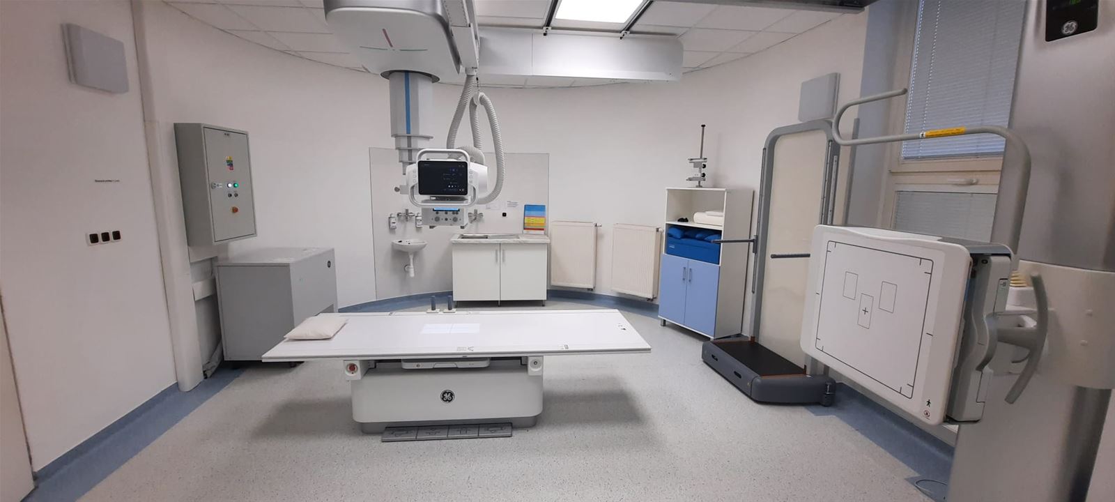
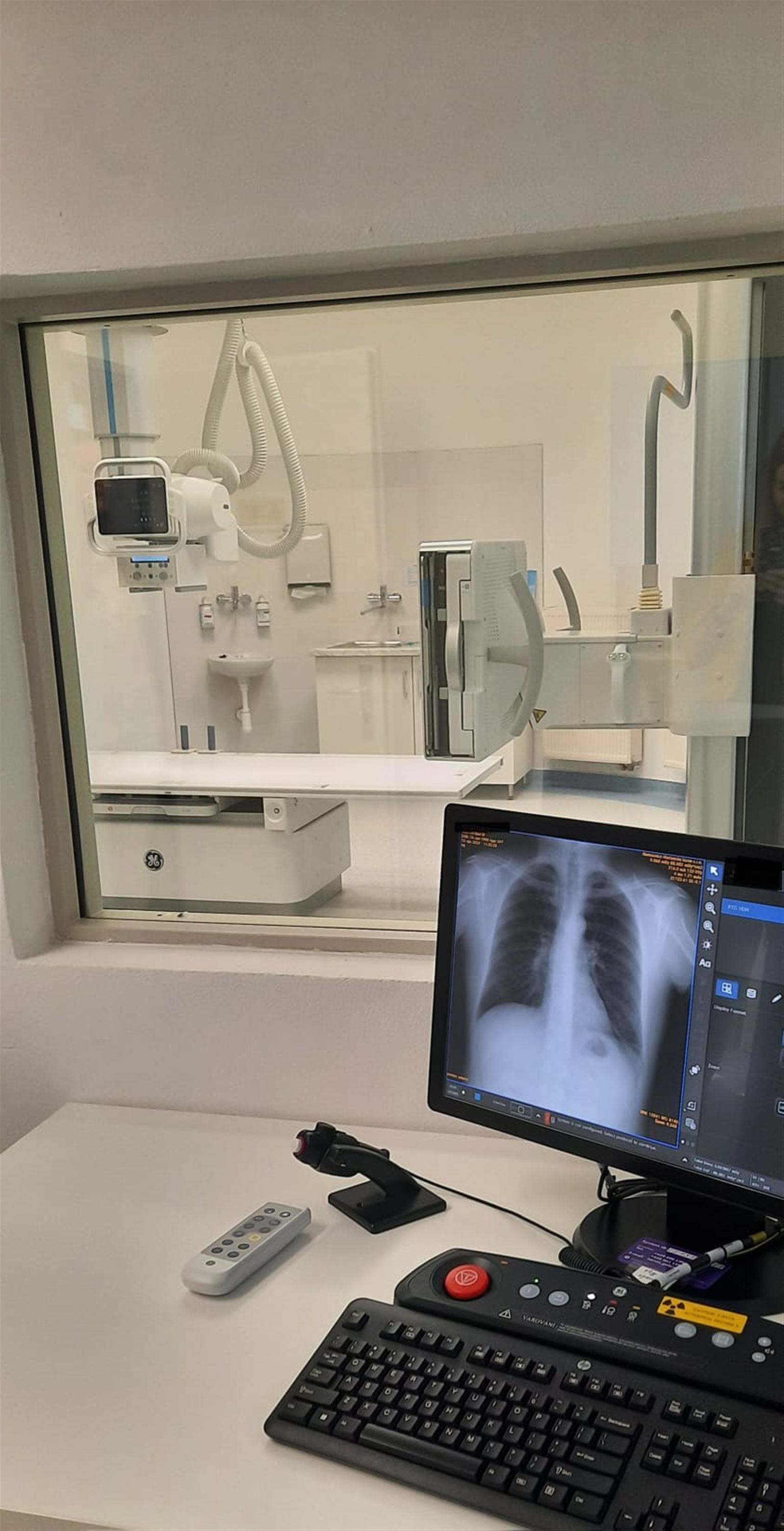
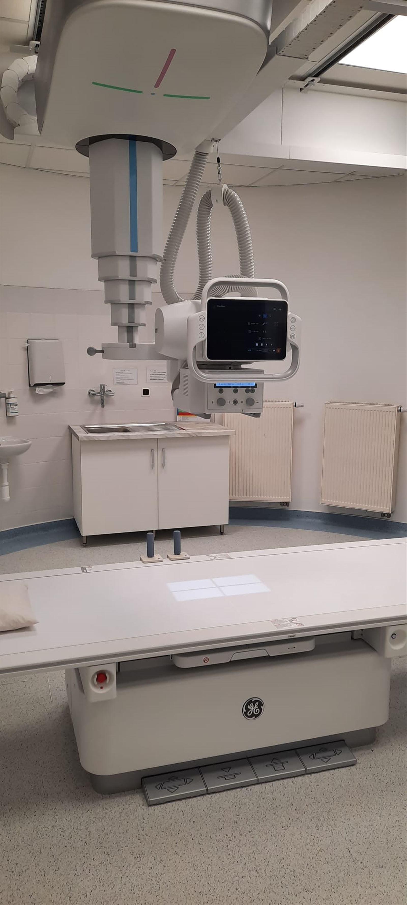
Instrumentation:
- device GE Definium Tempo Pro Philips by direct digitization - detectors Grip Sticker for FlashPad HD43x43. The load capacity of the examination table is max. 350 kg.
- skiagraphic device MP 15 with indirect digitization CR system Fuji Capsule XLII.
CT – computed tomography
Appointment time:
Monday - Friday: 7:00 - 14:00
Principle of the method:
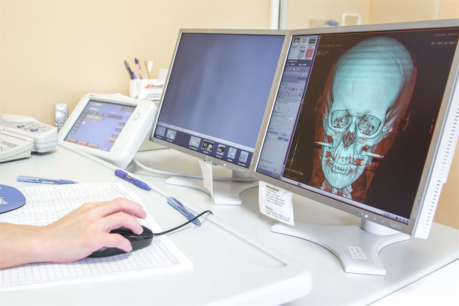
Computed multidetective tomography is a modern imaging method that uses X-rays in a special way to image individual parts of the body in thin layers. It is used for the diagnosis of a wide range of acute and chronic diseases. In order to visualize vessels, but also the structure of parenchymal organs and the characteristics of pathological lesions, it is necessary to administer iodine contrast liquid into a vein during the examination. Due to the relatively higher radiation load, CT is usually indicated only as a supplement in the case of unclear results on an ultrasound examination or a classic X-ray image. The disadvantage of this examination is the higher dose of ionizing radiation, therefore CT is performed only if there is a clear medical reason for the examination.
The examination must be ordered in advance! Acute cases (patients with a serious clinical condition) will be examined on a priority basis after prior phone call agreement between the attending physician and the CT staff. The order of the patients is decided by the doctor of the CT department.
The last patient will be admitted for examination no later than 30 minutes before the end of working hours.
Getting an apointment:
You can order an examination by phone 353 474 214 or in person. All patients who have a valid request form from a general practitioner or specialist can order an examination.
Examinations performed:
Brain CT, PND CT, Pyramidal CT (HRCT)
CT spine according to specification: C, Th, LS, CT SI joints
CT of the skeleton: e.g. skull including facial skeleton, shoulder, elbow, pelvis, knee, ankle...
CT of the neck
CT of the lungs and mediastinum, CT of the esophagus, CT angiography of the lungs, CT angiography of the thoracic aorta
CT of the abdomen and pelvis, CT angiography of the abdominal aorta, CT angiography of the renal arteries
CT of the limbs, CT angiography of the lower limbs
CT virtual colonoscopy – used to diagnose colorectal cancer, used after an incomplete or unfeasible optical colonoscopy
CT enterography (enteroclysis) – shows loops of the small intestine and is currently one of the best examinations of the small intestine. Most important pathological conditions of the intestines are well examined in loops of intestines properly filled with Mannitol.
Computed tomography without intravenous contrast can detect trauma, bleeding, stones in the urinary tract, sinus infections and herniated discs. Detections of these diseases are most common with CT without contrast material.
With the insertion of a contrast liquid into a vein, in addition to the above-mentioned diseases, various inflammatory and tumor conditions can also be diagnosed.
Preparation for a CT scan:
The patient fills in and signs consent to the examination on the form: "Informed consent - CT examination - computed tomography". Patients who are not scheduled to receive a contrast agent (native examination) do not need to fill in questions about allergies (inside the double sheet).
No special preparation is required before a CT scan without the application of a contrast agent.
Preparation for CT with intravenous contrast is here:
CT scan contraindications:
Native examination (i.e. examination without intravenous administration of contrast material) - a relative contraindication is pregnancy (for pregnant women, it can only be performed in the event of a threat to life). It is also not advised to examine patients with a CT shortly after an X-ray examination of the alimentary canal (e.g. X-ray with the act of swallowing, stomach...), as the barium substance from these examinations could invalidate the CT image. Just wait a few days or use laxatives.
We do not perform examinations with intravenous iodinated contrast agents with patients allergic to iodinated contrast agents, in cases of pregnancy, in patients with untreated uncorrected hyperthyroidism (increased thyroid gland function), and pheochromocytoma. For patients with renal insufficiency, we only perform the examination after approval by a nephrologist. For patients with asthma or a severe form of polyvalent allergy, the examination is only performed after premedication with Prednisone, which will be provided by the referring physician (for more see Preparation before application of iodine...), or with the assistance of an anesthetist
Limitations of the method:
- Obesity (carrying capacity of the CT device: 205 kg, the diameter of the gantry opening is also limiting)
- Claustrophobia
Risks:
X-ray radiation belongs to ionizing radiation. During it's life on Earth, the human body is constantly exposed to other types of ionizing radiation (cosmic radiation, radon...). Therefore, humans have developed very effective mechanisms to defend against potential radiation damage. Nevertheless, there is a small probability that even small doses can cause tumor growth or genetic damage in the body or in the irradiated fetus, which will manifest itself in the offspring. Tumors appear in the population for a variety of reasons, and a specific cause cannot usually be determined. The potential risk from medical radiation is incomparably lower in terms of probability than other risks that we encounter in everyday life and that we normally accept (traffic accidents, etc.). The used medical radiation alone cannot cause an immediate change in your state of health. During the examination, the X-ray radiation dose is as low as possible while maintaining the image quality.
The total radiation dose depends on the type of examination and the size of the examined area, but in general it can be said that the dose during a CT examination is noticeably higher than with a regular X-ray image, it is comparable to the dose of an examination with multiple images (e.g. an X-ray examination of the stomach or intestines)
Intravenous application of iodinated contrast material can have, just like any medicinal product, less serious or serious side effects or complications. For more information, see Informed consent - CT-computed tomography examination
Examination result:
Medical examination descriptions are usually drawn up on the day of the examination, no later than 3 working days after the examination, in the case of the so-called: STATIM examination (urgent medical examination), the descriptions are drawn up by the doctor immediately after the end of the examination. We perform STATIM description only until 14:00
Instrumentation: Bright Speed Elite 16-coil multi-detector fully digitized instrument (GE Medical), OptiVantage DH digital, remote-controlled injection system is used to apply contrast material
Appointment time
X-ray:
Monday - Friday: 7.00 – 15.30 (STATIM DESCRIPTION ONLY TILL 13.00)
CT, SONO
Monday -Friday: 7.00 – 14.00
EMERGENCY X-RAY
Monday - Friday: 15.30 – 21.00
Saturday - Sunday: 7.00 – 21.00
Contact information:
Phone number: +420 354 474 214